Technology Transfer
Imaging with very high energy electron
The DESY CMS group is developing a novel tomographic technique using an electron beam with energies from several hundreds megaelectronvolt to a gigaelectronvolt. The electrons transversing a target are deflected by multiple Coulomb scattering, and the angular distribution depends on the atomic number of the traversed material. The trajectory of the electrons is reconstructed using a pixel beam telescope with sensor planes situated in front of and behind the target. As a result of the high penetration depth of high-energy electrons, this technique has the potential to overcome challenges of conventional computed tomographies using X-ray or proton, regarding samples of high-Z materials or with large material budgets.
We visualized the inner structure of a test sample using a multi-GeV electrons delivered by the DESY test beam facility. The angular distribution of the reconstructed tracks in a target was used to create a sinogram for 3D image reconstruction of the sample. The picture shows a single slice of the reconstructed image of the DESY logo sample made of lead and aluminum.
We are currently working on a image reconstruction technique based on iterative methods and simulations for systematic effects estimation and development to the medical applications.
Compact Muon Solenoid Experiment
Imaging with very high energy electron
The DESY CMS group is developing a novel tomographic technique using an electron beam with energies from several hundreds megaelectronvolt to a gigaelectronvolt. The electrons transversing a target are deflected by multiple Coulomb scattering, and the angular distribution depends on the atomic number of the traversed material. The trajectory of the electrons is reconstructed using a pixel beam telescope with sensor planes situated in front of and behind the target. As a result of the high penetration depth of high-energy electrons, this technique has the potential to overcome challenges of conventional computed tomographies using X-ray or proton, regarding samples of high-Z materials or with large material budgets.
We visualized the inner structure of a test sample using a multi-GeV electrons delivered by the DESY test beam facility. The angular distribution of the reconstructed tracks in a target was used to create a sinogram for 3D image reconstruction of the sample. The picture shows a single slice of the reconstructed image of the DESY logo sample made of lead and aluminum.
We are currently working on a image reconstruction technique based on iterative methods and simulations for systematic effects estimation and development to the medical applications.
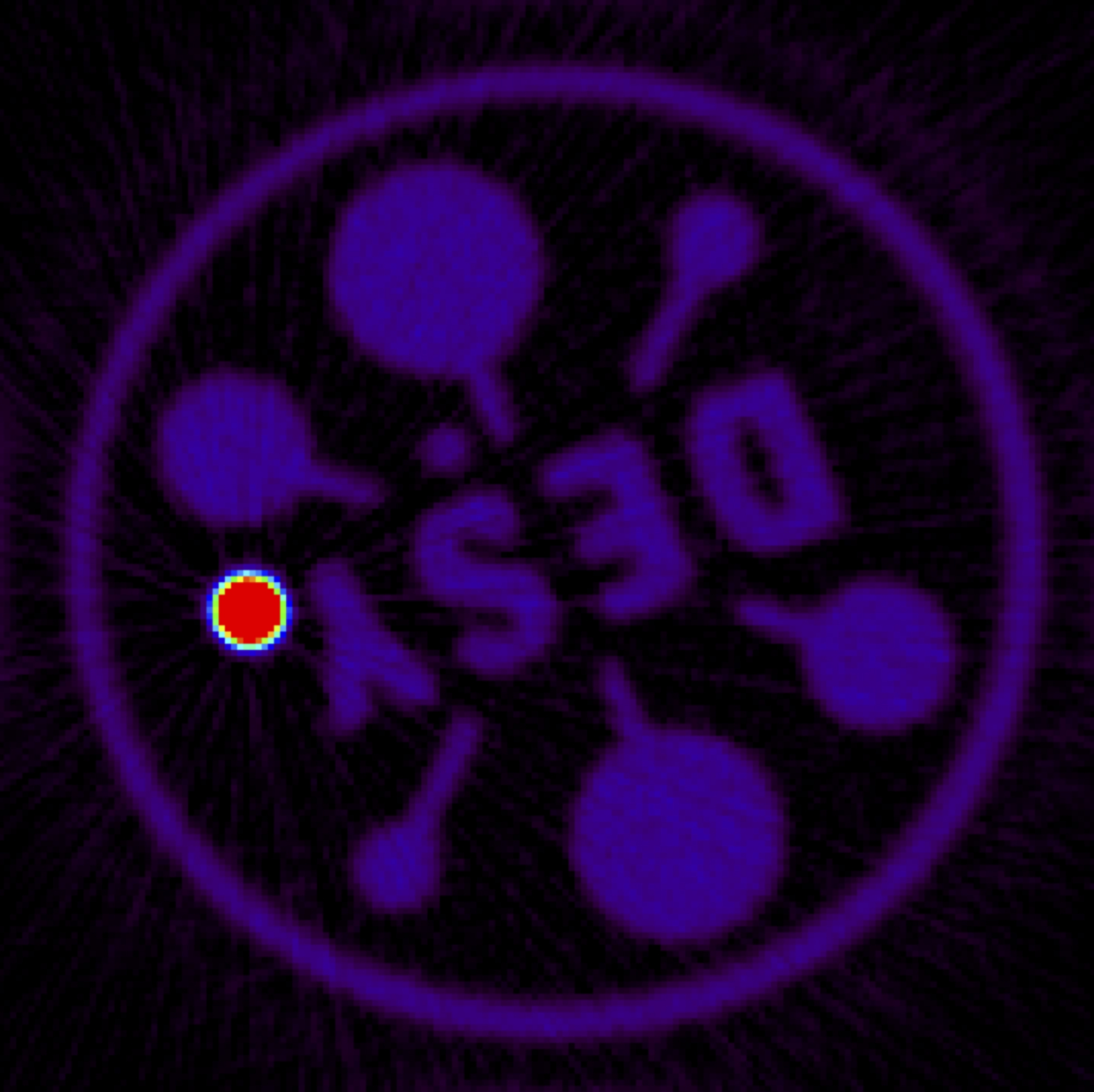
Reconstructed image of a DESY-logo phantom, which is made of an iron stick and plastic.